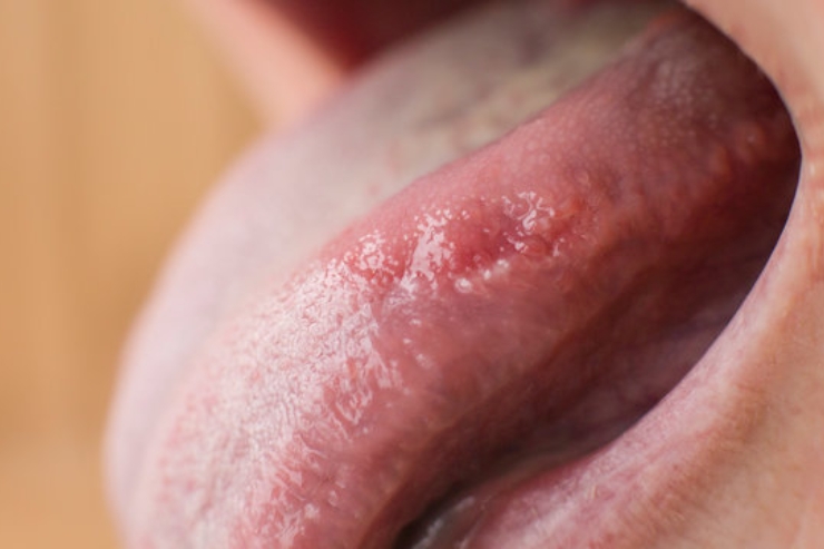
Laser excision of oral leukoplakia and erythroplakia
Laser Precision for Healthy Mucosa
Oral leukoplakia and erythroplakia are precancerous lesions—white or red patches, respectively—on the mucosal surfaces of the mouth. Laser excision uses focused beams (often CO₂ or diode lasers) to vaporize or sharply delineate these lesions with microscopic precision, minimizing damage to surrounding healthy tissue.
Personalized Treatment Plan:
Dr. Singhavi performs a detailed oral examination and maps each lesion’s extent with toluidine blue staining or autofluorescence guidance. Under local or light sedation, he adjusts laser settings for depth of penetration and tissue type, ensuring complete removal of dysplastic areas. The procedure is typically done on an outpatient basis. Postoperatively, he prescribes a tailored regimen of antimicrobial mouth rinses, topical steroids, and dietary modifications to support mucosal healing. Follow-up visits every 3–6 months include clinical inspection and repeat imaging if needed.
Benefits :
Laser excision under Dr. Singhavi’s care delivers clean margins with minimal bleeding, swelling, and postoperative discomfort. The precise energy delivery seals small blood vessels and nerve endings, accelerating healing and reducing pain. Patients appreciate the swift recovery—often returning to normal diet and speech within days. Regular surveillance combined with the minimally invasive laser approach lowers the risk of malignant transformation, ensures early detection of any recurrence, and maintains healthy oral mucosa for lasting comfort and confidence.