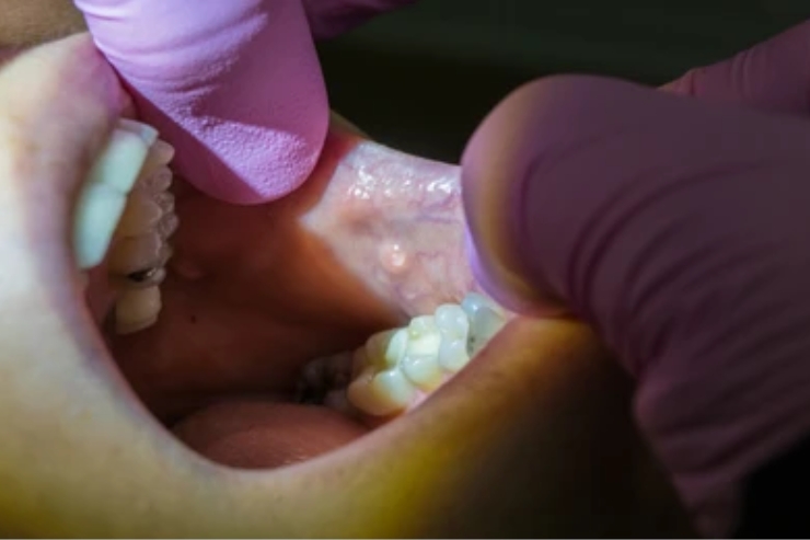
Sialendoscopy is a minimally invasive procedure to diagnose and treat obstructive salivary gland disorders—particularly stones (sialolithiasis) and ductal strictures—while preserving the gland. A miniature endoscope is introduced into the ductal system to visualize pathology, retrieve stones with micro-forceps, or dilate narrow segments.
Personalized Treatment Plan:
Dr. Singhavi orders ultrasound and non-contrast CT to map stone characteristics and duct anatomy. Under local anesthesia with sedation, he dilates the duct orifice and advances the sialendoscope under saline irrigation. Stones are captured with nitinol baskets or fragmented using a holmium laser when necessary. Balloon dilators address strictures. A temporary stent may be placed to maintain ductal patency. Post-procedure, patients follow a regimen of gland massage, hydration, and short-term antibiotics.
Benefits :
Sialendoscopy offers gland preservation, minimal discomfort, and no external scars. Procedures are typically outpatient, with most patients resuming normal activities the same day. By restoring natural salivary flow, it alleviates pain, swelling, and infection risk. Long-term outcomes include high success rates, reduced recurrence, and preserved gland function—ensuring sustained relief and excellent patient satisfaction.